fax:818-937-9182
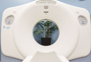
CT
A CT scanner uses digital geometry processing to generate a 3-dimensional (3D) image of the inside of an object. The 3D image is made after many 2-dimensional (2D) X-ray images are taken around a single axis of rotation – in other words, many pictures of the same area are taken from many angles and then placed together to produce a 3D image.
The Greek word tomos means “slice”, and the Greek word graphein means “write”.
Although CT is a useful tool for assisting diagnosis in medicine, it is a source of ionizing radiation and can cause cancer. The National Cancer Institute advises patients to discuss the risks and benefits of computerized tomography with their doctors.
How do CT scans work?
A CT scanner emits a series of narrow beams through the human body as it moves through an arc, unlike an X-ray machine which sends just one radiation beam. The final picture is far more detailed than an X-ray image.
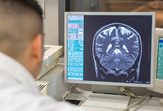
Inside the CT scanner there is an X-ray detector which can see hundreds of different levels of density. It can see tissues inside a solid organ. This data is transmitted to a computer, which builds up a 3D cross-sectional picture of the part of the body and displays it on the screen.
Sometimes a contrast dye is used because it shows up much more clearly on the screen. If a 3D image of the abdomen is required the patient may have to drink a barium meal. The barium appears white on the scan as it travels through the digestive system. If images lower down the body are required, such as the rectum, the patient may be given a barium enema. If blood vessel images are the target, the barium will be injected.
The accuracy and speed of CT scans may be improved with the application of spiral CT. The X-ray beam takes a spiral path during the scanning – it gathers continuous data with no gaps between images. For a spiral scan of the chest, for example, the patient will be asked to hold his/her breath for a few seconds.
When are CT scans used?
CT scanning is useful to get a very detailed 3D image of certain parts of the body, such as soft tissues, the pelvis, blood vessels, the lungs, the brain, abdomen, and bones.
It is often the preferred method of diagnosing many cancers, such as liver, lung, and pancreatic cancers. The image allows a doctor to confirm the presence of a tumor. The tumor’s size can be measured, plus its exact location, as well as to determine how much the tumor has affected nearby tissue.
A scan of the head can provide the doctor with important information about the brain – he/she may want to know whether there is any bleeding, swelling of the arteries, or tumors.
A CT scan will tell the doctor whether the patient has a tumor in his/her abdomen, and whether any internal organs in that area are swollen or inflamed. It will reveal whether there are lacerations of the spleen, kidneys or liver.
As a CT scan can detect abnormal tissue it is a useful device for planning areas for radiotherapy and biopsies.
A CT scan can also provide valuable data on the patient’s vascular condition. Vascular refers to blood flow. Many vascular conditions can lead to stroke, kidney failure, and even death. It can help a doctor assess bone diseases, bone density, and the state of the patient’s spine.
A CT scan can reveal vital data about injuries to the patient’s hands, feet and other skeletal structures – even small bones can be seen clearly, as well as their surrounding tissue.
Back To Blog
MAKE AN APPOINTMENT TODAY!
Please fill out the information below to request an appointment at our center.